Medical diagnostics is an ever-evolving field, and new advancements are continuously emerging to revolutionize how we diagnose and treat cardiovascular diseases. One of the most valuable tools in this field is 2D echocardiography, a non-invasive imaging technique that provides real-time, detailed images of the heart’s structure and function. As the demand for skilled echocardiographers increases, specialized training programs, such as fellowships in 2D echocardiography, become more important.
Understanding 2D Echocardiography
Before delving into the significance of fellowship programs, it’s crucial to grasp the fundamentals of 2D echocardiography. Unlike traditional imaging methods, such as X-rays or CT scans, echocardiography utilizes sound waves to create images of the heart. By emitting high-frequency sound waves and analyzing their reflections off cardiac structures, this technique produces detailed two-dimensional images of the heart chambers, valves, and major blood vessels.
2D echocardiography, also known as two-dimensional echocardiography, is a vital imaging modality used in cardiology to visualize the heart’s structure and function in real time. It provides detailed, high-resolution images of the heart in motion, allowing clinicians to assess cardiac anatomy, chamber dimensions, valve function, and overall cardiac performance.
There are several types of 2D echocardiography techniques, each offering unique advantages in specific clinical scenarios. Here are some common types:
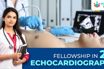
1. Transthoracic Echocardiography (TTE)
- Transthoracic echocardiography is the most commonly performed type of echocardiography.
- It involves placing the ultrasound transducer on the chest wall to obtain images of the heart.
- TTE provides a comprehensive assessment of cardiac structure and function, including evaluation of chamber size, wall thickness, valve morphology, and systolic and diastolic function.
- This technique is non-invasive, well-tolerated by patients, and can be performed at the bedside or in the outpatient setting.
2. Transesophageal Echocardiography (TEE)
- Transesophageal echocardiography involves inserting a specialized ultrasound probe into the esophagus to obtain images of the heart from behind.
- TEE provides clearer and more detailed images of cardiac structures, particularly the posterior cardiac structures that may be difficult to visualize with TTE.
- It is often used in patients with suboptimal TTE images, intraoperative monitoring during cardiac surgery, assessment of cardiac sources of embolism, and evaluation of prosthetic heart valves.
- TEE requires sedation and is typically performed in a controlled environment such as the cardiac catheterization laboratory or operating room.
3. Stress Echocardiography
- Stress echocardiography combines echocardiographic imaging with physical exercise (e.g., treadmill exercise or pharmacological stress) to assess cardiac function under conditions of increased workload.
- It is used to evaluate myocardial ischemia, viability, and contractile reserve in patients with suspected coronary artery disease.
- Stress echocardiography can help identify regions of the myocardium that do not receive adequate blood flow during stress, indicating areas of potential coronary artery obstruction.
4. Contrast Echocardiography
- Contrast echocardiography involves the administration of contrast agents (microbubbles) to enhance ultrasound imaging of the heart.
- Contrast agents improve endocardial border delineation and enhance visualization of cardiac structures, particularly in patients with suboptimal image quality.
- This technique is valuable for assessing myocardial perfusion, detecting intracardiac shunts, and evaluating cardiac masses or thrombi.
5. Fetal Echocardiography
- Fetal echocardiography is a specialized form of echocardiography used to evaluate the structure and function of the fetal heart during pregnancy.
- It plays a crucial role in the prenatal diagnosis of congenital heart defects, allowing for early detection and appropriate management of cardiac anomalies.
- Fetal echocardiography requires specialized training and expertise in fetal imaging and prenatal cardiology.
Each type of 2D echocardiography has its indications, advantages, and limitations. The selection of the appropriate echocardiographic technique depends on the clinical scenario, the information needed, and the patient’s condition. By leveraging these diverse imaging modalities, clinicians can obtain comprehensive insights into cardiac anatomy and function, guiding optimal patient management and improving outcomes in cardiovascular care.
The Role of Fellowship Programs
While basic training in cardiology equips physicians with essential skills in echocardiography interpretation, fellowship programs provide specialized training that goes beyond the basics. A fellowship in 2D echocardiography offers a structured curriculum designed to enhance expertise in image acquisition, interpretation, and clinical decision-making.
These programs typically span one to two years and provide hands-on experience under the guidance of experienced echocardiographers.
Key Components of Fellowship Training
- Advanced Imaging Techniques: Fellows gain proficiency in advanced imaging modalities, such as Doppler echocardiography, strain imaging, and three-dimensional echocardiography. These techniques allow for a comprehensive assessment of cardiac structure and function, aiding in the diagnosis and management of various cardiovascular conditions.
- Clinical Correlation: Beyond technical skills, fellowship programs emphasize the integration of echocardiographic findings into clinical practice. Fellows learn to correlate imaging data with patient history, physical examination findings, and other diagnostic tests to formulate accurate diagnoses and treatment plans.
- Research and Innovation: Many fellowship programs offer opportunities for fellows to engage in research projects aimed at advancing the field of echocardiography. By participating in research endeavors, fellows contribute to the development of new imaging techniques, diagnostic algorithms, and therapeutic strategies.
Advantages of Fellowship Training
- Expertise and Competence: Completion of a fellowship in 2D echocardiography instills fellows with a high level of expertise and confidence in performing and interpreting echocardiographic studies. This specialized training enhances diagnostic accuracy and improves patient outcomes.
- Career Advancement: Fellowship training opens doors to diverse career opportunities in academic institutions, private practice, and research settings. Echocardiographers with fellowship training are highly sought after for their specialized skills and knowledge.
- Networking and Collaboration: Fellowship programs foster collaboration and networking among fellows, faculty members, and experts in the field. These connections provide valuable mentorship, professional support, and opportunities for ongoing learning and growth.
Fellowship training in 2D echocardiography is a crucial component of cardiovascular medicine. It plays an essential role in advancing clinical expertise, improving diagnostic accuracy, and driving innovation in this field. By providing a comprehensive curriculum that focuses on advanced imaging techniques, clinical correlation, and research, these programs prepare fellows to become the next generation of highly skilled echocardiographers.
As the importance of echocardiography continues to grow, fellowship training remains instrumental in shaping the future of cardiovascular imaging and enhancing patient outcomes.