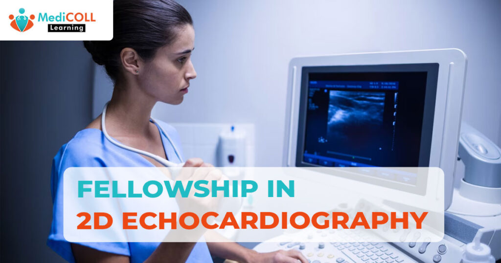
In the dynamic field of cardiovascular medicine, the role of advanced imaging techniques has become indispensable for accurate diagnosis and effective patient care. Among these, 2D echocardiography stands out as a powerful tool that allows clinicians to visualize the heart in real-time, providing valuable insights into its structure and function. The pursuit of excellence in this domain often involves engaging in a fellowship in 2D echocardiography, a journey that combines specialized training, hands-on experience, and collaboration with experts in the field. The field of medical imaging has undergone remarkable advancements in recent years, providing healthcare professionals with powerful tools to assess and diagnose various conditions.
One such essential technique is 2D echocardiography, a non-invasive imaging method that offers a detailed view of the heart’s structure and function. In this blog post, delve into the intricacies of cardiography, exploring its principles, applications, and significance in the realm of cardiology. Two-dimensional echocardiography, is a diagnostic imaging technique that utilizes ultrasound waves to generate real-time, two-dimensional images of the heart. It provides a dynamic and detailed visualization of the heart’s chambers, valves, and surrounding structures, allowing healthcare professionals to assess cardiac anatomy and function without the need for invasive procedures. Fellowship in 2D echocardiography is a specialized training program designed to equip healthcare professionals with the knowledge and skills necessary to become experts in the field.
Principle: The basic principle behind 2D echocardiography involves the transmission of high-frequency sound waves (ultrasound) into the chest, which then bounce off the heart’s structures and return as echoes. These echoes are detected by a transducer, a handheld device that emits and receives ultrasound waves. The collected data is processed in real-time to create a two-dimensional image of the heart on a monitor.
Key Components of 2D Echocardiography
- Transducer: The transducer is a crucial component that emits ultrasound waves and captures the returning echoes. It is placed on the patient’s chest at specific locations to obtain different views of the heart.
- Ultrasound Gel: Before placing the transducer, a gel is applied to the patient’s chest. This gel enhances the transmission of ultrasound waves and ensures better contact between the transducer and the skin.
- Echocardiographic Views: Different views, such as the parasternal, apical, and subcostal views, allow for a comprehensive assessment of the heart’s structures and function. These views provide valuable information about the chambers, valves, and blood flow within the heart.
Applications of 2D Echocardiography
- Structural Assessment: 2D echocardiography is instrumental in evaluating the size, shape, and thickness of the heart’s chambers, as well as the integrity of the valves and surrounding structures.
- Functional Analysis: It provides dynamic information about the heart’s contraction and relaxation, enabling the assessment of cardiac function, ejection fraction, and valve motion.
- Diagnosis of Cardiovascular Conditions: 2D echocardiography is widely used in the diagnosis of various cardiovascular conditions, including heart valve disorders, cardiomyopathies, congenital heart defects, and pericardial diseases.
- Monitoring and Follow-up: Healthcare professionals use 2D echocardiography to monitor patients with known cardiac conditions, track the progression of diseases, and assess the effectiveness of interventions.
Advantages of 2D Echocardiography
- Non-Invasive: Unlike invasive procedures such as cardiac catheterization, 2D echocardiography does not require penetration of the skin or insertion of instruments into the body.
- Real-Time Imaging: It provides immediate and dynamic images, allowing healthcare professionals to observe the heart’s structures and function in real-time.
- Safety: Ultrasound waves used in 2D echocardiography are considered safe and do not involve exposure to ionizing radiation, making it suitable for repeated examinations.
Key Components of a Fellowship in 2D Echocardiography:
- Didactic Education: Fellows undergo comprehensive didactic education covering the principles of ultrasound physics, cardiac anatomy, and the interpretation of echocardiographic findings. This foundational knowledge forms the basis for advanced imaging techniques.
- Hands-On Training: Practical experience is a cornerstone of the fellowship. Fellows participate in supervised echocardiography sessions, learning to perform and optimize imaging studies. This hands-on training allows them to develop proficiency in acquiring high-quality images and understanding the technical aspects of the equipment.
- Case Reviews and Interpretation: Fellows engage in the interpretation of a diverse range of echocardiographic cases under the guidance of experienced mentors. This aspect of the fellowship helps them refine their diagnostic skills and learn to correlate imaging findings with clinical data.
- Research Opportunities: Many fellowships include a research component, encouraging fellows to contribute to the advancement of knowledge in the field. Research projects may focus on refining imaging techniques, exploring novel applications of 2D echocardiography, or investigating the impact of imaging on patient outcomes.
Collaboration and Networking
Fellowship in 2D echocardiography provides a unique opportunity for fellows to collaborate with multidisciplinary teams. Interaction with cardiologists, cardiac surgeons, and other healthcare professionals enhances the fellows’ understanding of the broader clinical context in which echocardiography plays a crucial role.
Challenges and Rewards
While the journey through a 2D echocardiography fellowship may present challenges, such as the steep learning curve and the need for meticulous attention to detail, the rewards are equally significant. Graduates emerge as skilled echocardiographers, capable of making precise diagnoses and contributing to advancements in cardiovascular care.
Fellowship in 2D echocardiography is a transformative experience that propels healthcare professionals into the forefront of cardiovascular imaging. It combines theoretical knowledge with hands-on training, fostering expertise and proficiency in the nuanced world of cardiac ultrasound. As these fellowship graduates contribute to the field, their impact extends beyond individual patient care, shaping the future of cardiovascular medicine through innovation and excellence.


0 Comments